
Gynecological Exams / Tests
Mammography
No device at the clinic
This sentence is the first working rule of this page. SoftCare is a women’s health clinic for examinations, pelvic ultrasound, breast ultrasound and taking laboratory samples. There is no mammography unit, no compression device and no mammography reporting unit here. We do not write “we do it too”.
The doctor looks at screening age, family history and any palpable finding. If mammography is indicated, the patient is referred in writing to the partner centre. The centre takes the images and returns the report with a BI-RADS category. If needed, ultrasound or biopsy is the job of that centre or of the unit the patient is referred to. SoftCare keeps the report and tells you the next examination interval and any open question. The day of the exam, the dose and the device brand are not the subject of this page.
Screening and symptoms are not the same door
Screening is for catching cancer early in a woman without symptoms. Diagnostic mammography is requested for a palpable lump, bloody discharge from a single duct, a new nipple retraction or a skin change. The second does not wait for age. Under thirty, the first imaging is usually ultrasound; if doubt remains, mammography is added to that file and done at the centre.
| Situation | What is done |
|---|---|
| No symptoms, 40–69, average risk | National programme: mammography every two years |
| No symptoms, average risk, ACOG framework | Start at 40; interval of 1 or 2 years, chosen together |
| Palpable hardness | No waiting for age; ultrasound and mammography if needed |
| Bloody discharge from a single duct | Diagnostic work-up, referral to the centre |
| Cancer at a young age in mother or sister | Risk is assessed separately; interval and MRI may change |
| Pregnant, palpable lump | Ultrasound first; if mammography is unavoidable, at the centre with the abdomen shielded |
What the exam is like
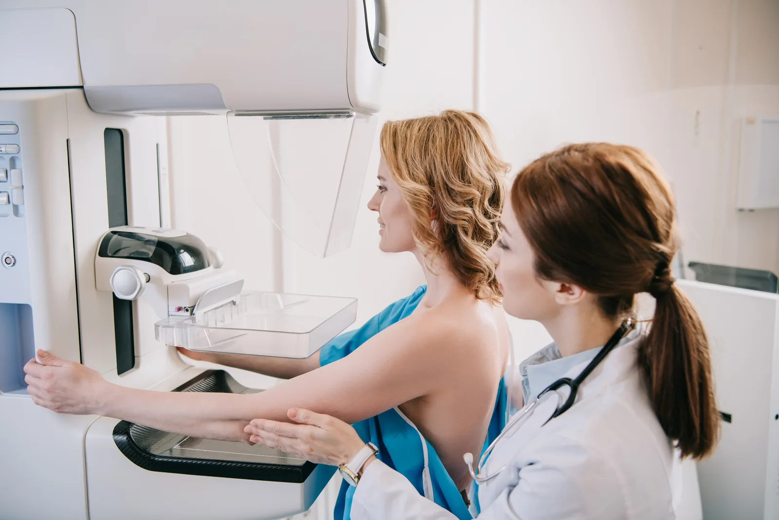
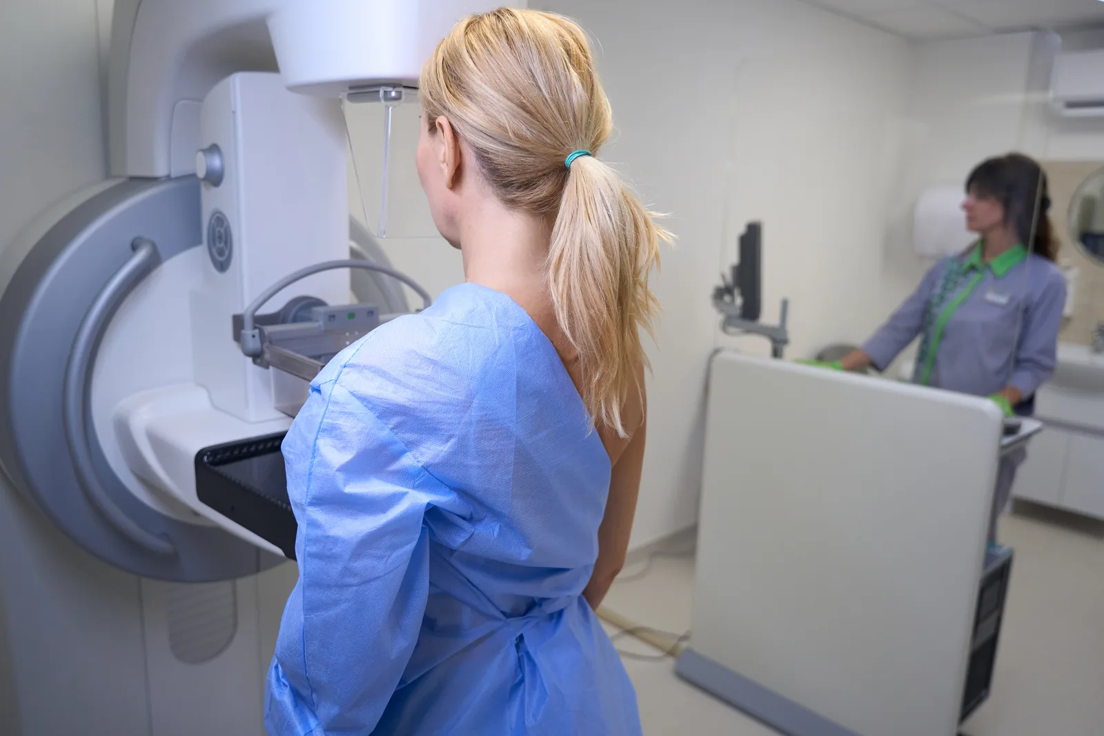
The breast is compressed between two plates for a few seconds. Compression lowers the dose and spreads the tissue apart. For most women, the pain is brief. The week after the period ends may be more comfortable; for screening, that week is not required. Do not apply deodorant or talc to the armpits on the day of the exam; they can show up as specks. The exam is done standing, and at least two views are taken of each breast.
The dose is in the range of a few chest X-rays; in a woman at average risk, the benefit of early diagnosis from screening is weighed against this dose. In pregnancy, screening is postponed. If there are symptoms, ultrasound comes first. If mammography is unavoidable, the abdomen is covered and the decision is made with the centre and the doctor.
Silicone or saline implants do not prevent the exam. Extra views that move the implant aside are needed. An implant can lower the quality of screening; this is not a reason to skip the exam. Implant rupture is the job of ultrasound and magnetic resonance imaging, not a promise of mammography.

Dense breasts
Density means that glandular tissue looks more prominent than fatty tissue on mammography. Four categories are used: fatty, scattered fibroglandular, heterogeneously dense, extremely dense. In heterogeneously and extremely dense breasts, some cancers can be masked. This does not cancel mammography. The ACR discusses supplemental screening based on risk and density. Ultrasound or magnetic resonance imaging can be added. Additional imaging is not put in place of mammography. SoftCare does not use density as an excuse to say “we covered it with ultrasound” at the clinic.
How to read the report
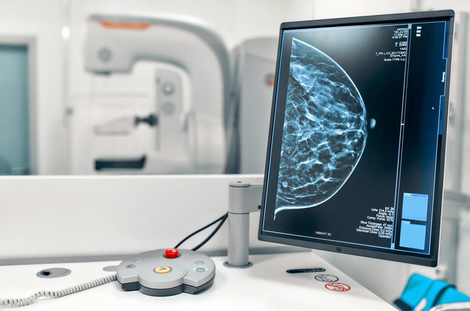
The mammography result comes back with a BI-RADS category. This is not a stage. It tells you the next step.
| Category | What it means | Next step |
|---|---|---|
| 0 | Incomplete assessment | Additional images or ultrasound |
| 1 | No significant finding | Age-appropriate screening |
| 2 | Benign appearance | Routine follow-up |
| 3 | Probably benign | Short-interval check |
| 4 | Suspicious | Biopsy is discussed |
| 5 | Highly suspicious | Biopsy |
| 6 | Known cancer | Treatment follow-up |
Category 0 is not a sentence to fear. The images were not enough. Category 3 does not mean forgetting; a check-up whose date is missed loses its meaning. The biopsy decision reads the category in the report and the examination together. Once SoftCare receives the report, we explain this distinction; we do not repeat the imaging here.
National programme and referral from the clinic
The Ministry of Health’s national breast screening programme offers women at average risk a mammogram every two years between ages 40 and 69. It can be done at KETEM (cancer early-diagnosis centres), healthy living centres and affiliated screening units. This route is the door to free screening. SoftCare does not replace these centres and does not book their appointments either.
If there is a family history, a previous suspicious report or a palpable finding, a written referral is made to the partner imaging centre. What we ask of the patient is to bring her old images and reports. Without comparison, a new image is not closed as “clear”. When the result is out, the doctor tells you the category, whether further imaging is needed and the next date.
Who needs a different interval
Average risk is the name of the framework used when there is no breast or ovarian cancer at a young age in the family, no known gene change and no history of radiotherapy to the chest wall. If one of these is present, screening before 40 and with magnetic resonance imaging may be discussed. Genetic testing is not the job of this page. Risk is assessed at the examination, not stuck on paper as a “high risk” label.
After seventy, a woman is outside the standard range of the national programme. Other illnesses and life expectancy decide whether screening continues or stops. Age alone does not forbid the exam.
What it does not show
Mammography does not catch every cancer. In dense breasts, sensitivity drops. A cancer found between screenings does not mean the previous image was wrong. Benign calcification and a suspicious cluster are not always told apart on the first image; additional imaging and biopsy exist for that. It does not give a hormone result. It does not certify that an implant is intact. It may not always pick out a simple cyst that ultrasound shows.
Our approach at SoftCare
Mammography is not done here. Examination, breast ultrasound and history stay at the clinic. A woman of screening age without symptoms is referred to the national programme or the partner centre. A palpable lump, bloody discharge and skin change are investigated regardless of age; if mammography is needed, she goes to the centre through the same door. The report comes back, the category is read, and at the clinic missing images and biopsy are not closed with “ultrasound is enough”.
Frequently asked questions
Is mammography done at SoftCare?
No. There is no device. A patient who needs one is referred to a partner imaging centre.
At what age does it start?
In Türkiye, at average risk, the national programme covers ages 40–69, every two years. ACOG says screening should start at 40.
Is it painful?
The compression is brief. It may be more comfortable after your period.
Isn’t ultrasound enough?
Not for screening. It is not put in place of mammography.
Does a palpable lump wait for age?
No.
Do dense breasts cancel the exam?
No. Additional imaging is discussed; it does not stop the mammography decision.
Are old images needed?
Yes. Without comparison, the new report remains incomplete.
Is category 0 cancer?
No. The assessment is incomplete.
Listing these sources does not mean SoftCare performed the imaging or wrote those texts.
References
- Meme Kanseri Tarama Programı Ulusal Standartları. T.C. Sağlık Bakanlığı, Halk Sağlığı Genel Müdürlüğü, 2026.
- ACOG Clinical Practice Update: Age to Initiate Routine Breast Cancer Screening. American College of Obstetricians and Gynecologists (ACOG). Obstetrics & Gynecology 145(1), 2025.
- Practice Bulletin No. 179: Breast Cancer Risk Assessment and Screening in Average-Risk Women. American College of Obstetricians and Gynecologists (ACOG). Obstetrics & Gynecology 130(1), 2017.
- ACR Appropriateness Criteria: Supplemental Breast Cancer Screening Based on Breast Density, 2024 Update. American College of Radiology. Journal of the American College of Radiology 22(5), 2025.
- ACR BI-RADS Atlas: Mammography. American College of Radiology, 2013.
Medically reviewed by Op. Dr. Şükrü Sanus, Obstetrician & Gynecologist · Last medical review: