
Gynecological Exams / Tests
Ovarian Cysts

Types
Functional cysts:
- Follicular cyst: Forms when the follicle keeps growing because ovulation did not occur. Usually disappears within 3–8 weeks.
- Corpus luteum cyst: Forms when fluid or blood collects inside the corpus luteum after ovulation. May cause a late period or mild pain. It can also be seen in early pregnancy.
Other cysts:
- Dermoid cyst (mature cystic teratoma): Develops from embryonic cells. It may contain fat, hair, teeth or skin tissue. Most are benign.
- Cystadenoma: Develops from cells on the surface of the ovary. It can be serous or mucinous. It may grow; most are benign.
- Endometrioma (chocolate cyst): Forms as a result of endometriosis. It contains dark blood. It may be linked to painful periods and difficulty getting pregnant.
- Polycystic ovary: An appearance of many small follicles. This is a separate condition and is not covered on this page.
Malignant cysts are rare. Risk is assessed using the ultrasound appearance, age and tumour markers.
Symptoms
Most cysts cause no symptoms. If there are symptoms:
- Pain in the groin on one or both sides (dull or sharp)
- A feeling of fullness or pressure in the lower abdomen
- Bloating
- Pain during sex (especially deep)
- Irregular periods or bleeding between periods
- Frequent urination or constipation (with large cysts)
Sudden severe pain on one side, nausea, vomiting or feeling faint suggest torsion or rupture. In this case, emergency assessment is needed.
Diagnosis
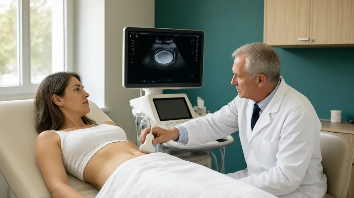
Diagnosis is made by gynaecological examination and ultrasound. Transvaginal ultrasound shows the cyst's size, wall thickness, septa, solid areas and blood flow. Simple, thin-walled cysts with a single chamber are usually considered functional.
If the appearance is suspicious (solid areas, thick septa, irregular borders, ascites), MRI or tumour markers (CA-125) may be discussed. CA-125 alone cannot make a diagnosis; it can also rise with endometriosis and inflammation. If pregnancy is possible, a pregnancy test is done first. The IOTA and O-RADS classifications are used to assess risk on ultrasound.

Follow-up and treatment
During the reproductive years, simple-looking, small (usually under 5 cm) cysts without symptoms are usually monitored with a follow-up ultrasound after 6–8 weeks. Most disappear on their own. After menopause, simple cysts are assessed more carefully; further tests are considered for cysts over 3–5 cm or with a suspicious appearance.
Reasons for surgery:
- Suspicious ultrasound appearance
- A cyst that causes symptoms or is growing
- Suspected torsion or rupture
- Large cystadenoma or dermoid
- Endometrioma and a wish to become pregnant
Surgery is usually done as a laparoscopic cystectomy. Every effort is made to preserve the ovary. If malignancy is suspected, oncological surgery is planned.
Complications
- Torsion: Twisting of the ovary around itself. It causes sudden pain, nausea and vomiting. It requires emergency surgery.
- Rupture: Bursting of the cyst. It can cause sudden pain and sometimes internal bleeding.
- Malignancy: Rare. A suspicious appearance requires further assessment.
In the IOTA5 study, cysts with a benign ultrasound appearance had a low risk of malignancy, torsion and rupture over 2 years of follow-up. This supports monitoring of simple cysts.
When to seek emergency care
- Sudden severe pain in the groin on one side
- Pain together with nausea, vomiting or fainting
- Severe pelvic pain with fever
- Pain or bleeding when pregnancy is possible
- Rapidly increasing abdominal swelling or pain spreading to the shoulder
Our approach at SoftCare
At SoftCare Clinic, ovarian cyst assessment begins with a gynaecological examination and ultrasound. The cyst's size, appearance and any symptoms are recorded. For small cysts with a simple appearance, a follow-up ultrasound is planned. If the appearance is suspicious, the cyst is large or there are emergency signs, further assessment or referral is arranged. If endometrioma is suspected, it is addressed together with the endometriosis page. If pregnancy is possible, a pregnancy test is done first. In cases that need surgery, referral to a suitable centre is arranged.
Appointments: +90 212 241 30 10 · softcareclinic.com
References
- 1. American College of Obstetricians and Gynecologists (ACOG). Ovarian Cysts. FAQ.
- 2. Mayo Clinic. Ovarian cysts – Symptoms and causes.
- 3. Society of Radiologists in Ultrasound (SRU). Consensus on Simple Adnexal Cysts Follow-up (2019 update).
- 4. International Ovarian Tumor Analysis (IOTA). IOTA5 study – Conservative management of adnexal masses. Lancet Oncology, 2019.
- 5. American College of Radiology. O-RADS US v2022.
- 6. European Society of Gynaecological Oncology / ISUOG / IOTA / ESGE Consensus on preoperative assessment of adnexal masses.
- 7. Merck Manual Professional Edition. Ovarian Cysts.
- Note: Follow-up intervals, surgical indications and risk assessment are updated by the physician according to current guidelines. This reference list is for information only.
Listing a source does not mean SoftCare wrote that text.
Medically reviewed by Op. Dr. Şükrü Sanus, Obstetrician & Gynecologist · Last medical review: