
Gynecological Exams / Tests
Uterine Fibroids (Myoma)
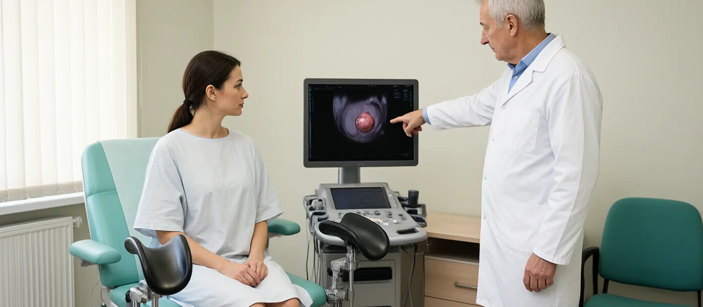
Location (FIGO classification)
The FIGO system describes how a fibroid relates to the uterine cavity and the outer surface of the uterus:
- Type 0–2: Submucosal (grows into the uterine cavity). More often linked to bleeding and difficulty getting pregnant.
- Type 3–4: Intramural (within the uterine wall).
- Type 5–7: Subserosal (grows outward). May press on the bladder or bowel.
- Type 8: Other locations (cervix, etc.).
Location affects the choice of treatment. Hysteroscopic removal is more suitable for submucosal fibroids.
Symptoms
- Heavy or prolonged menstrual bleeding (may lead to anaemia)
- Spotting between periods
- Pelvic pressure or fullness
- Frequent urination or constipation (with large fibroids)
- Pain during sex
- Lower abdominal or lower back pain
- Rarely, difficulty getting pregnant or recurrent miscarriage (especially submucosal)
Having no symptoms does not mean a fibroid is harmless; however, small fibroids without symptoms are usually monitored.
Diagnosis
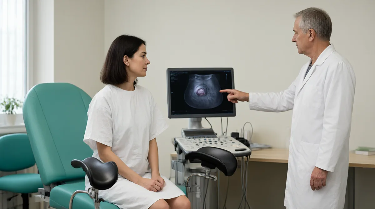
Diagnosis is made by gynaecological examination and ultrasound. Transvaginal ultrasound shows the size, number and location of fibroids. If a submucosal fibroid is suspected, sonohysterography or hysteroscopy may be needed. For large or multiple fibroids, MRI shows the boundaries more clearly.
The differential diagnosis includes adenomyosis, ovarian cysts and, rarely, sarcoma. If there is anaemia, a complete blood count is ordered. If pregnancy is possible, a pregnancy test is done first.
Treatment options
Small fibroids without symptoms can be monitored. If there are symptoms, the options are:
Medication:
- Tranexamic acid or NSAIDs (to control bleeding)
- Hormonal contraceptives or progestogens
- GnRH agonists or antagonists (short-term shrinkage)
- Selective progesterone receptor modulators (in some countries)
Procedures:
- Uterine artery embolisation: The blood vessels that feed the fibroid are blocked. Suitable for multiple fibroids. Not preferred if you plan a pregnancy.
- MR-guided focused ultrasound (MRgFUS) or radiofrequency ablation: In selected cases.
Surgery:
- Myomectomy: Removal of the fibroid while keeping the uterus. Hysteroscopic for submucosal fibroids; laparoscopic or open for others. Preferred if you plan a pregnancy.
- Hysterectomy: A definitive solution if symptoms are severe and you do not wish to keep your uterus.
Treatment is chosen according to age, severity of symptoms, number and location of fibroids, pregnancy plans and personal preference. The ACOG 2021 guideline supports uterus-sparing options.
Pregnancy and fertility
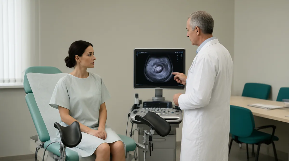
Submucosal fibroids can make it harder to get pregnant and can increase the risk of miscarriage. The effect of intramural and subserosal fibroids is less clear. If you plan a pregnancy, myomectomy is usually preferred over embolisation. Fibroids may grow during pregnancy; most cause no problems. Rarely, they may be linked to pain or preterm birth.
When to see a doctor
- Heavy bleeding, clots or signs of anaemia
- Pelvic pressure, frequent urination or constipation
- Rapid growth or growth after menopause
- Difficulty getting pregnant or recurrent miscarriage
- Sudden severe pain (suspected torsion or degeneration)
Our approach at SoftCare
At SoftCare Clinic, fibroid assessment begins with a gynaecological examination and ultrasound. Size, number and FIGO location are recorded. If there is anaemia, a complete blood count is ordered. For small fibroids without symptoms, monitoring is planned. If there are symptoms, medication, embolisation or surgical options are discussed. If you plan a pregnancy, myomectomy takes priority. In cases that need hysterectomy or advanced procedures, referral to a suitable centre is arranged. If there is abnormal bleeding, assessment of the endometrium is considered.
Appointments: +90 212 241 30 10 · softcareclinic.com
References
- 1. American College of Obstetricians and Gynecologists (ACOG). Management of Symptomatic Uterine Leiomyomas. Practice Bulletin No. 228, June 2021 (reaffirmed 2025).
- 2. Stewart EA, Laughlin-Tommaso SK. Uterine Fibroids. N Engl J Med. 2024;391:1721–1733.
- 3. International Federation of Gynecology and Obstetrics (FIGO). Leiomyoma subclassification system.
- 4. Munro MG, Critchley HOD, Fraser IS. The FIGO classification of causes of abnormal uterine bleeding. Int J Gynaecol Obstet. 2011;113:3–13.
- 5. American Society for Reproductive Medicine (ASRM). Removal of myomas in asymptomatic patients to improve fertility and/or reduce miscarriage rate.
- 6. Merck Manual Professional Edition. Uterine Fibroids.
- 7. UpToDate. Uterine fibroids (leiomyomas): Epidemiology, clinical features, diagnosis, and natural history.
- Note: Treatment options, suitability for embolisation and surgical indications are updated by the physician according to current guidelines. This reference list is for information only.
Listing a source does not mean SoftCare wrote that text.
Medically reviewed by Op. Dr. Şükrü Sanus, Obstetrician & Gynecologist · Last medical review: